Cellular and molecular biomechanics, microscopy

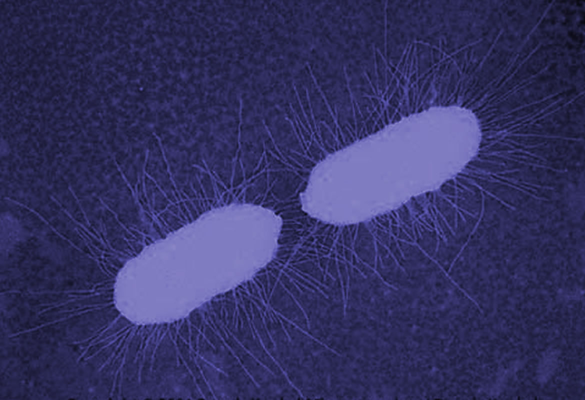
Bacteria have appendices similar to hair named pili, which allow the adhesion of bacteria cells to human cells in flow conditions. In E. coli, this adhesion promotes the generation of infections such as cystitis. In previous research, it has been shown that pili can extend like elastic strings to limit the effect of big forces caused by the fluid flow in the adhesive end of pili. In our previous projects we have shown that the molecule FimH, in charge of the adhesion between pili and the surface, presents amazing adhesive properties. Besides, we have evidenced that the pili has a remarkable property: it can have a greater adhesion under tension than without tension. This counter-intuitive property is called catch bond, quite similar to the finger traps found in kid’s toys.

In a collaboration project with the Mechanical Engineering department, we want to run simulations and experiments on multiple levels to understand the way in which the fluid, pili and FimH properties affect bacterial adhesion.


Light sheet fluorescence microscopy (lsfm) applied to development studies in zebrafish

The study of the mechanisms underlying both neural development and functioning in vivo is today possible thanks to light sheet fluorescence microscopy (LSFM) techniques, such as Single Plane Illumination Microscopy (SPIM). In this project, we develop a methodology to image developing zebrafish embryos using the SPIM. This allows us to identify neural structures expressing an extracellular matrix protein, F-spondin, and to characterize migration patterns of optic tectum (Tec) neurons. We image zebrafish embryos (transgenic line spon1b:GFP) from 24 to 70 hours post-fertilization (hpf), and we have identified possible primordia of olfactory bulbs (OB) and habenular (Hb) progenitors at 24 hpf. Additionally, we apply image processing and analysis to obtain single cell trajectories and the morphology, to determine the mechanisms that might be important during optic tectum development.

Super resolution microscopy with 3b analysis

Beyond the optical limit –where it does’nt matter how good a microscope is- hide minuscule structures inside the cell. To be able to watch them and understand them better, super resolution techniques have been developed. These make use of the turning on and off of fluorescent molecules to produce images with nanometric scale detail. We work on the implementation of one of these techniques (3B analysis microscopy) in our lab. The following image (false color) shows the quality that can be reached. Frame 1 shows the fluorescent sample as seen on the microscope with a 100x objective. The scale (2000nm) is shown in white and the region of interest analyzed is shown in yellow. Frame 2 is a digital zoom of the region of interest shown in figure 1. Frame 3 shows the reconstruction in super resolution of the region of interest. The difference between the resolution of frames 2 and 3 (which show the same object) is notorious. This reconstruction in super resolution was obtained from 940 frames that were analyzed computationally during 1100 minutes.

Jorge Madrid y Christian Poveda

FaLang translation system by Faboba
  • Cra 1 Nº 18A - 12
    Edificio Q | Ext 3783
    Bogotá - Colombia

  • Edificio IP | Ext 2730
    +(571) 339 49 99
    +(571) 339 49 49

Universidad de los Andes | Vigilada Mineducación
Reconocimiento como Universidad: Decreto 1297 del 30 de mayo de 1964.
Reconocimiento personería jurídica: Resolución 28 del 23 de febrero de 1949 Minjusticia.
Carrera 1 18A-10, Bloque IP, Bogotá - Colombia. A.A. 4976-12340.
Teléfono +571 3324500. Fax +571 3324516
Universidad de los Andes - Facultad de Ciencias - Departamento de física.